Introduction
The term ‘paleoradiology’, coined by Notman, refers to the study of bioarchaeological materials using such medical imaging techniques (Chhem and Brothwell 2008,1). This paper provides an in depth account of the issues present in current paleoradiological research. Several commonly occurring issues have been identified, namely regarding sample sizes, anatomical positioning of human remains, poor image quality produced by thick CT slices, conservation, issues associated with bog bodies, and concerns with equipment, and issues of interpretation.
Sample sizes
A great deal of paleoradiological research is conducted with a case study approach (eg. Asingh and Lynnerup 2007; Gostner et al 2011; Panzer et al 2017; etc.), which has been widely criticised. Elliott (2018,6) acknowledges that some researchers have put effort into increasing their sample sizes, but the majority of research still focuses on case studies. Nelson and Wade (2015,942) are extremely critical of this approach, describing it as ‘highly limiting’, noting that such research can make it difficult to identify regional, temporal, and social trends.
Some issues with the case study method, as presented by Hodkinson and Hodkinson (2001,10-11), are that the results are not generalisable, and are easily dismissable by those who disagree with the results. For researchers like Wade and Nelson (2013,4198), who stress the value of avoiding ‘pan-Egyptian stereotypes’, it is obvious that these are serious issues. Their finding that transnasal excerebration was much less common in Egyptian mummification than suggested by classical depictions (Nelson and Wade 2015,943), would not have been as dramatic a discovery if it had been based on an individual mummification, examined using a case study method. Using a larger and more representative sample ensures that findings are more widely generalisable and harder to dismiss.
Anatomical positioning
CT scanners are designed for the medical investigation of living human beings, who are typically able to move themselves into a position allowing them to fit inside the scanner, which is obviously not the case when dealing with human remains. A common issue occurring with investigations of bog bodies, mummies, and other examples of archaeological human remains, such as the Pompeii casts (Lazer 2017,9), is awkward anatomical positioning which makes it difficult for remains to be placed into a CT scanner.
For instance, Grauballe Man’s right elbow did not fit into the scan field of the CT scanner, so was not recorded in the CT image (Asingh and Lynnerup 2007,95). The positioning of the Tyrolean Iceman’s left arm meant the body had to be placed into the scanner once head first and again feet first, in order to scan the entire body (Gostner et al 2011,3426). The positioning of the body may also have caused the ulnae to appear shorter on CT scans (Gostner et al 2011,3429). In the case of the Llullaillaco mummies, scan techniques had to be adapted to the anatomic position of each mummy (Previgliano et al 2003,1474). As seen in the cases of the Tyrolean Iceman and the Llullaillaco mummies, adaptations can be made to overcome anatomical issues, although these adaptations represent a compromise rather than a solution to these issues.
CT slice thickness
The thickness of CT ‘slices’, the intervals between each x-ray, hugely impacts image quality, and thicker CT slices can lead to structure misinterpretation (Lynnerup 2009,366). Modern CT scanners can achieve slices of less than 1mm thick, compared to the several millimetre slice thickness of earlier scanners (Lynnerup 2009,366). This means that modern CT images are much higher quality than their predecessors, generally speaking. However, scans of the Llullaillaco mummies conducted by Previgliano et al (2003,1474) have a slice thickness of 5mm, obtained at 5mm intervals. CT working sessions were limited to 20 minutes to prevent thawing and to conserve the bodies, however this produces an impression of the structure of the bodies with a limited accuracy.

Issues associated with bog bodies
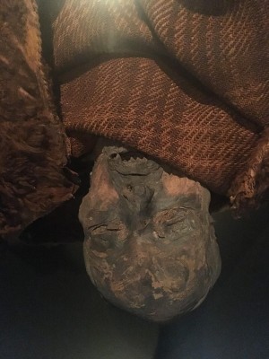
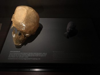
There are several challenges presented by bog bodies when using medical imaging. Due to the taphonomic influences of the bog environment, calcium content of bone is reduced, causing the tissue to become softer and less dense (Lynnerup 2010,444). Soft tissues, such as skin and tendons, seem to increase in density within the bog environment (Asingh and Lynnerup 2007,99; Lynnerup 2010,445). These factors combine to result in poor visualisation of the internal structures of bog bodies when the clinical range of Hounsfield Units are applied to the scans (Lynnerup 2009,367; Lynnerup 2010,444). In the case of Grauballe Man, the high radiodensity of the soft tissues and the low radiodensity of the skeletal tissues meant the structures were difficult to differentiate, and the CT scans had to be carefully manually segmented using MIMICS software (Asingh and Lynnerup 2007,114). One of the femurs of Lindow II shows considerable loss of bone density due to the acidity of the peat bog, and the Huldremose bog body also displays poor bone density (Chhem and Brothwell 2008,67-68).
(Photograph, M. Schlanker 2018)
(Photograph, M. Schlanker 2018)
Another issue presented by bog bodies is the methods of ‘conservation’ used soon after the bodies were discovered often make it difficult to form any meaningful observations on more recent scans. When the body of Grauballe Man was reappraised using x-ray and CT in the early 2000s, no signs of disease was found but the filling material inserted into the body during the 1950s conservation process could have obscured any fractures or signs of disease (Asingh and Lynnerup 2007,109). This means the findings of the reappraisal are not as reliable, as there may have been evidence of damage or pathologies that were simply obscured. There were also no remains of internal organs found in the abdomen, and what appears to be blackboard erasers in the chest cavity (Asingh and Lynnerup 2007,118-119). This of course means the internal organs could not be investigated paleoradiologically as part of the body. Methods of conserving bog bodies in the 1950s also frequently involved dehydration, which makes the use of MRI more of a challenge when investigating the remains (Elliott 2018,9; Shin et al 2010,329).
James Elliott (2018,9) believes bog bodies are not ideal candidates for paleoradiology, namely due to a great degree of post-mortem disfiguration (both before and after excavation), and the de-mineralisation of bone. I am inclined to disagree with Elliott, given that research by Villa and Lynnerup (2012,127) has already provided a set of Hounsfield Unit ranges for tissues mummified under peat bog conditions, with the aim that the knowledge of typical HU ranges will help to prevent the misinterpretation of CT images and improve their clarity without extensive manual post-processing.
Issues associated with equipment and interpretation
There are issues associated with the use of hospital equipment for paleoradiology. Radiologists used to working with living patients are not trained in dealing with bioarchaeological materials, and there is the issue of taking resources away from medical cases (Chhem and Brothwell 2008,15). Anthropology labs are unlikely to have as advanced equipment, including CT scanners, compared with hospitals, and anthropology lab equipment is often designed for smaller specimens and therefore cannot be used to investigate mummies or large bones (Chhem and Brothwell 2008,15).
Conclusion
A range of issues are prominent within current paleoradiological research. These include issues relating to the case study approach, such as results not being generalisable and therefore easy to dismiss, although researchers such as Nelson and Wade (2015) have put concerted effort into using larger sample sizes. The anatomical positioning of human remains can make it impossible for the body to be examined using CT and can also result in distorted radiographic images, and issues presented by using thicker CT ‘slices’ when scanning, usually to preserve the integrity of frozen remains, resulting in limited anatomical accuracy. Researchers must also deal with conversational concerns and liaise with the museums and sites at which the remains are housed, which can slow, limit, or prevent paleoradiological investigations. Another major issue is advanced equipment, such as CT scanners, which are only available at hospitals.
Bog bodies present several unique issues in the field of paleoradiological research, leading some, including Elliott (2018, 9) to argue they are not ideal candidates for this form of investigation. Early conservation techniques, involving filling material and rehydration, make radiographic reappraisal and the use of MRI more challenging, and changes in bone density and soft tissues lead to poor visualisation. However, Villa and Lynnerup (2012) have established a range of Hounsfield Units for bog bodies, aiding anatomical visualisation.
Recent work appears to be reducing the impact of paleoradiological issues. Villa and Lynnerup’s (2012) HU ranges allow for clearer radiographic images and less ambiguity in interpretation, Chhem and Brothwell (2008) call for more interaction between radiographers and archaeologists, and databases such as Nelson and Wade’s (2015) I.M.P.A.C.T. reduce the negative impact of the case study method. While there definite issues remain, these advancements mark positive developments in paleoradiology.


![Figure 1: Map of cemeteries studied in this essay. Porter, C, "Map of Cemeteries Used" [JPG map], Scale 1:100000, GB Overview [ geospatial data], Updated: 2019, Ordnance Survey (GB), Using: EDINA Digimap Ordnance Survey Service, <http://digimap.edina.ac.uk/>, Created: October 2019.](https://theposthole.org/sites/theposthole.org/files/styles/article_thumb/public/articles/454/fig%202.png?itok=cGHEw2KW)